varicocele
- related: Urology
- tags: #note
Varicoceles are caused by dilation of the testicular vein and pampiniform plexus. They are common, occurring in 15% of men. Scrotal examination reveals a left-sided (90%) scrotal mass with a “bag of worms” consistency that increases with standing and decreases while supine. This patient's findings are not consistent with varicocele, and examining the patient in both standing and supine positions is unlikely to support this diagnosis.
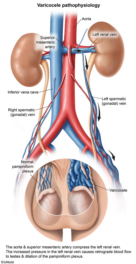
This patient's presentation suggests a varicocele, which is due to tortuous dilation of the pampiniform plexus of veins surrounding the spermatic cord and testis in the scrotum. Varicoceles occur in up to 20% of men (usually age 15-25); they are usually left sided, but up to 30% can be bilateral. The left spermatic (gonadal) vein enters the left renal vein at a right angle. The aorta and superior mesenteric artery can compress the left renal vein and increase intravascular pressure within the left renal and gonadal veins. This can cause incompetence of the valves, retrograde blood flow, and venous dilation.
Patients can be asymptomatic or develop a dull ache with standing. Examination usually shows a soft mass that is enlarged with standing and Valsalva maneuvers but diminishes when supine. Ultrasound can show retrograde venous flow and dilation of pampiniform plexus. Asymptomatic patients do not require treatment. Those with scrotal discomfort usually improve with analgesics (eg, nonsteroidal anti-inflammatory drugs) and/or scrotal support. Potential complications include infertility and testicular atrophy (due to increased scrotal temperature).
Because the right gonadal vein empties directly into the inferior vena cava, right-sided varicoceles are rare and can indicate inferior vena cava compression (eg, renal cell carcinoma) or obstruction (eg, thrombus). Sudden onset of left-sided varicocele that does not diminish in the recumbent position may also indicate obstruction. CT imaging is suggested in such cases but is otherwise not necessary.