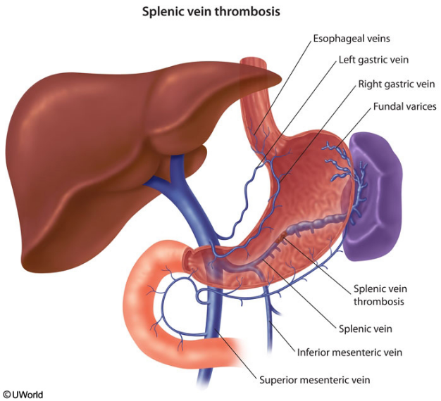
splenic vein thrombosis
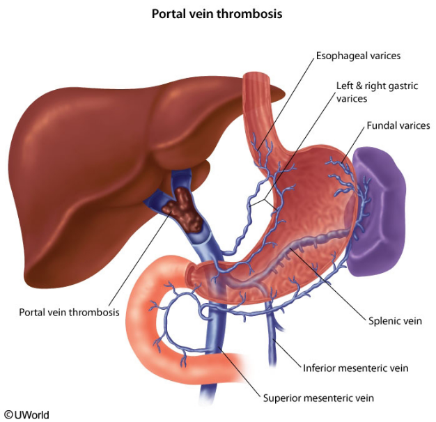
- related: GI, portal vein thrombosis
This patient presenting with gastrointestinal hemorrhage and isolated gastric varices in the setting of prior pancreatitis likely has splenic vein thrombosis (SVT). Most patients with SVT have a history of acute or chronic pancreatitis or pancreatic cancer. Thrombosis occurs as the splenic vein runs along the posterior surface of the pancreas and can become damaged or compressed due to pancreatic inflammation (or pancreatic masses/pseudocysts).
Patients with SVT are frequently asymptomatic; however, when symptoms do occur, the most common presentation is variceal bleeding. The isolated gastric varices, which are the hallmark of SVT, form when venous blood flow is redirected from the splenic vein to the collateral gastroepiploic system and short gastric veins. Patients with chronic SVT may also develop left-sided portal hypertension (due to isolated pressure increases in the splenic portion of the portal system), ascites, and congestive splenomegaly with associated features of hypersplenism (eg, anemia, thrombocytopenia).
The diagnosis of SVT can be confirmed directly with multiple imaging modalities, including a contrast-enhanced CT scan, Doppler ultrasonography, and MRI. It can also be confirmed indirectly with esophagogastroduodenoscopy by identifying isolated gastric varices. The standard treatment for patients with SVT and gastrointestinal hemorrhage is splenectomy.