immune complex mediated glomerulonephritis
- related: Nephrology
- tags: #nephrology
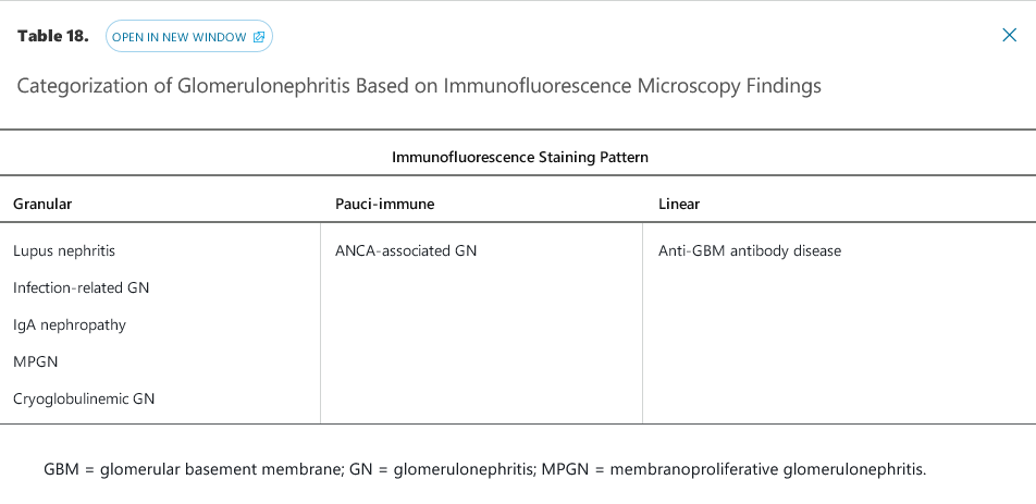
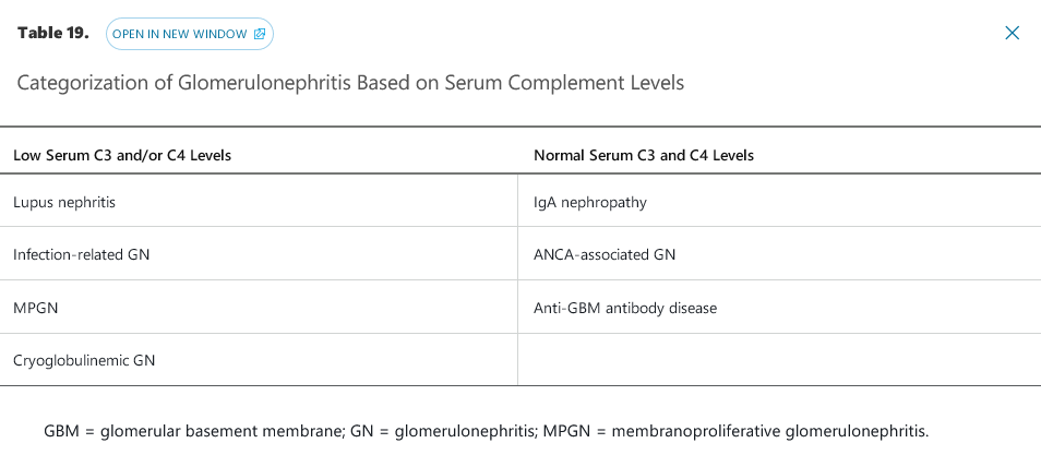
The glomerulonephritides with granular immunofluorescence microscopy staining patterns all fall under the umbrella category of immune complex–mediated glomerulonephritis, with antigen-antibody complexes depositing in the kidney at various locations and in various patterns. Immune complex deposition activates the classic complement pathway as a co-player in glomerular inflammation, injury, and, if unchecked, scarring. For this reason, most immune complex–mediated glomerulonephritides are associated with low levels of C3 and/or C4. IgA nephropathy, which usually presents with normal C3 and C4 levels, is an exception due to the chronic, gradual nature of the disease; complement consumption is at a rate slow enough for hepatic production to replace complement proteins. In rare hyper-acute cases of IgA nephropathy that fulfill criteria for RPGN, however, complement levels are usually depressed.


- cryoglobulinemic GN: associated with hepatitis C