hand fractures
A Colles fracture is another common fracture caused by falling onto an outstretched hand. It is a dorsally angulated or displaced distal radius fracture that is typically associated with visible angulation proximal to the wrist joint ("dinner fork deformity"). Lateral radiographs can confirm Colles fracture diagnosis.
Patients with Colles' fracture are also at risk for acute carpal tunnel syndrome. Acute management of Colles' fracture is dependent on the displacement and angulation of the fracture as well as any additional injuries. Most can be managed with conservative measures such as sugar tong splinting, with or without closed reduction. More severe injuries, especially those with significant displacement or angulation >15-20 degrees may require urgent orthopedic consultation for possible surgical intervention.
Scaphoid fracture
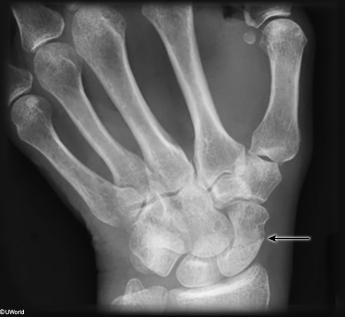
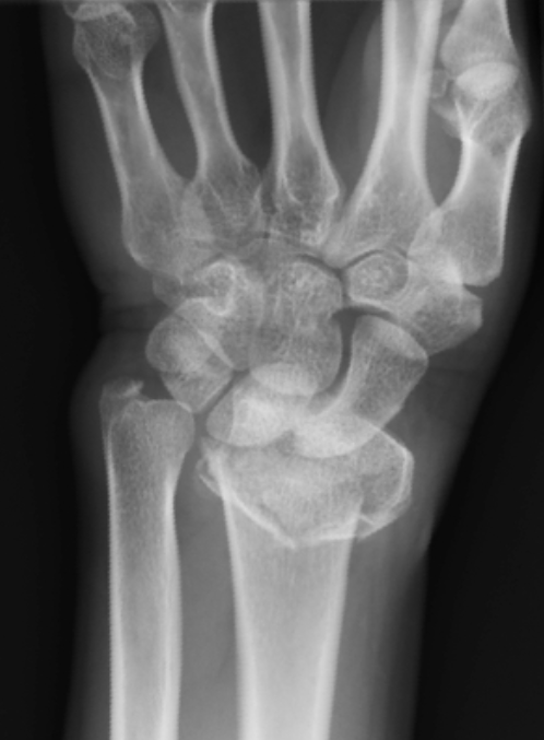
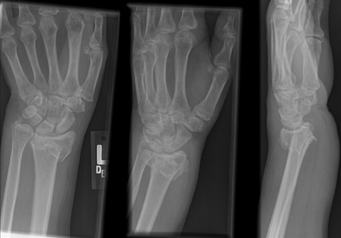
This patient fell onto his outstretched hand and has acute pain at the wrist and tenderness at the anatomic snuffbox. These are clinical features concerning for a scaphoid fracture, which is the most common carpal bone fracture and is typically caused by forceful dorsiflexion at the wrist. The scaphoid can be forced against the dorsal part of the distal radius, creating a fulcrum for injury. Patients can have swelling, decreased grip strength, pain and tenderness at the radial aspect of the wrist in the anatomic snuffbox, and minimally decreased range of motion (unless dislocated fracture is present).
The next step in management would be to confirm the diagnosis with x-rays of the wrist in full pronation and ulnar deviation to better expose the scaphoid. Initial x-rays can be negative if the fracture is compressed or minimally displaced.
Initial plain radiography of a scaphoid fracture may reveal the fracture but may appear normal if the fracture is compressed or minimally displaced. In light of the limited sensitivity of initial x-rays, patients with suspected scaphoid fracture should have additional imaging:
- MRI (or CT scan) of the wrist
- Repeat radiographs in 7-14 days
- Radioscintigraphy bone scan in 3-5 days
MRI is the most sensitive test and allows for immediate assessment of a fracture, but is subject to limited availability and high up-front costs. Alternatively, the patient may be placed in a thumb spica splint with repeat x-rays in 7-14 days, but this approach can lead to delayed diagnosis, unnecessary immobilization, and greater follow-up costs.
Patients with a confirmed scaphoid fracture should be referred to an orthopedic surgeon if they are found to have associated tilt of the lunate, a fracture displaced >1 mm, nonunion during follow-up, osteonecrosis, or scapholunate dissociation . Patients with a nondisplaced fracture who do not meet surgical indications should be placed in a short arm thumb spica cast; serial x-rays should be done in 2-week intervals to monitor healing.