child and elderly abuse
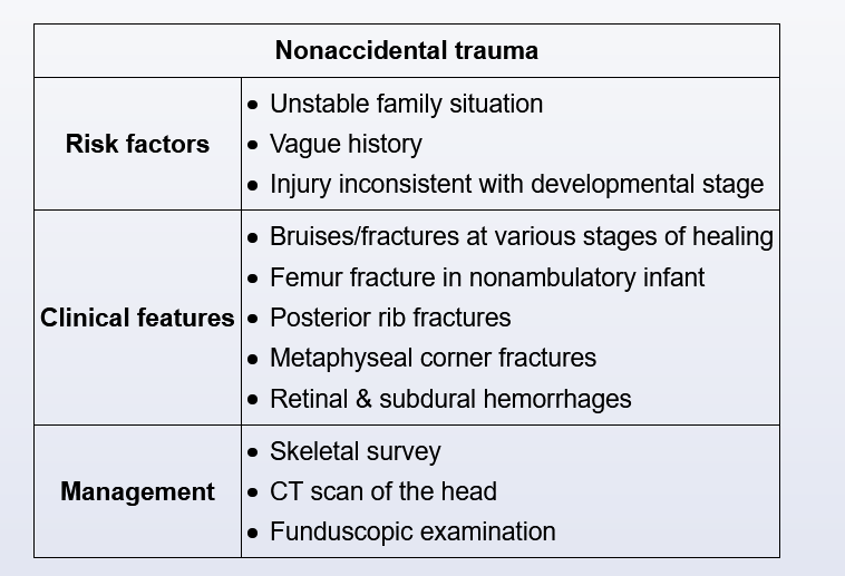
Nonaccidental trauma (NAT), or child abuse, occurs most frequently in infants age <1. Delayed presentation to medical care is typical, and red flags for abuse include a vague history of injury and a supposed mechanism that is inconsistent with the child's development. Siblings or pets are also commonly blamed for the injury.
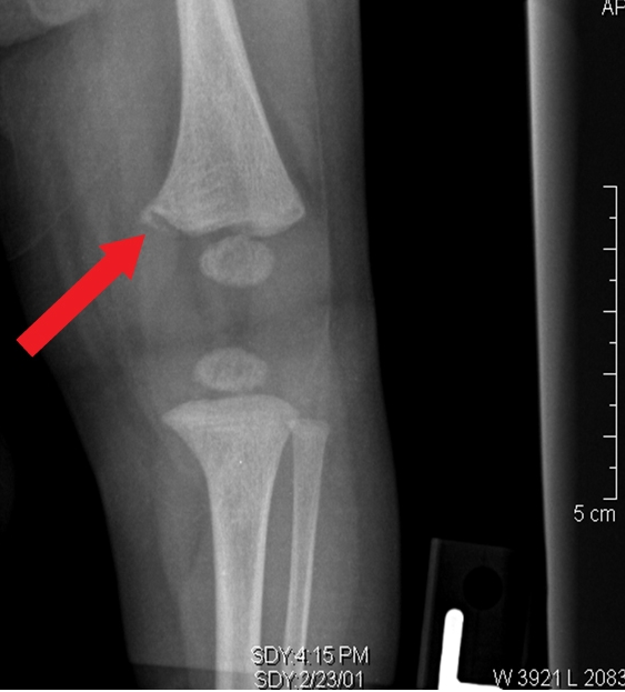
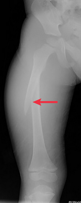
Certain fracture patterns are warning signs of abuse. A femur fracture in a nonambulating child, particularly a spiral fracture as seen in this patient, is suspicious for a twisting force on the thigh. The reported mechanism of injury in this case is unlikely, as most 18-month-olds can run but not jump off a couch. Furthermore, the sibling's weight is likely not sufficient for a direct fall to result in a fracture; if it were, a transverse fracture would be expected. In addition, the sibling's history of digital fractures is concerning for inflicted injury by door slamming. Other fractures that are virtually pathognomonic for NAT include posterior rib fractures, metaphyseal corner ("bucket-handle") fractures, and fractures at various stages of healing.
Given this patient's presentation and x-ray findings, a complete skeletal survey, fundoscopy, and CT scan of the head are necessary to evaluate for other fractures, retinal hemorrhages, and intracranial injury (eg, subdural hematoma).
Neurofibromatosis type I (NF1), an autosomal dominant disorder characterized by an increased tumor susceptibility (eg, neurofibromas, optic gliomas), presents with >6 café-au-lait macules. This patient's 3 small macules are normal. In addition, there is no suggestion of a positive family history for NF1.
Osteogenesis imperfecta, due to defective type 1 collagen, causes fractures, retinal hemorrhages, and bruising similar to NAT. However, patients typically have multiple fractures in the neonatal period and blue sclera, not present in this case.
Osteomalacia, soft bones due to decreased bone mineralization, can be caused by rickets (vitamin D deficiency) in children. Pathologic fractures are common, but epiphyseal widening, metaphyseal cupping, and osteopenia are expected on radiograph.
Metaphyseal corner: