Disorders of the Mucous Membranes
- related: Dermatology
- tags: #dermatology
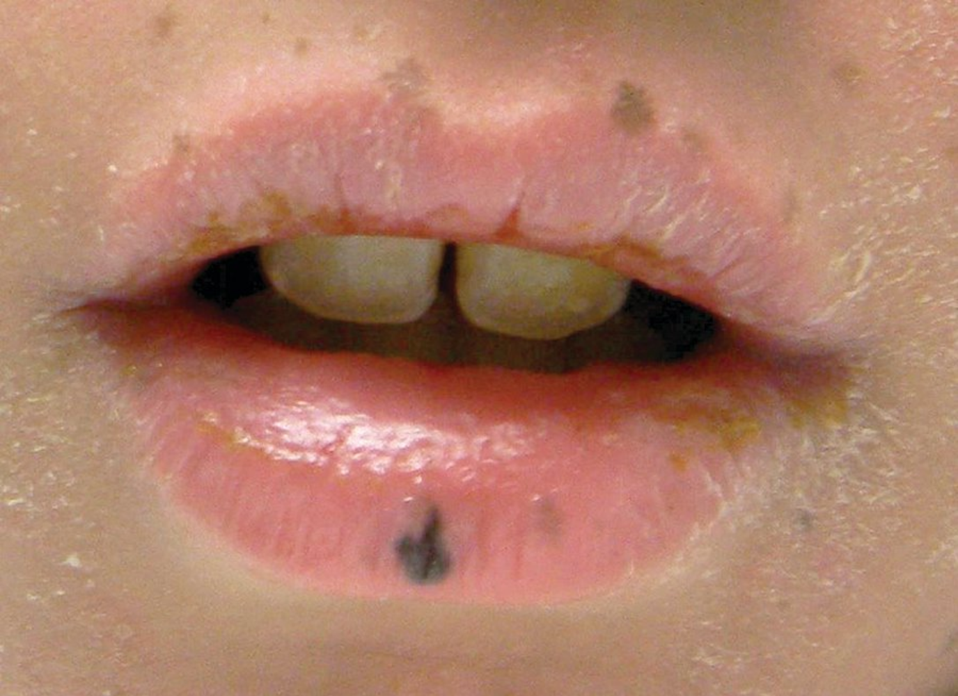
Melanotic Macule
A melanotic macule is a small, well-circumscribed, brown-to-black mucosal lesion. The most common location is the lower lip (Figure 149), but they may also be found on the gingival, buccal mucosa, or tongue, or within the genital mucosa. If large, papular, or not well-circumscribed, biopsy should be performed to rule out melanoma. Solitary lesions are the rule, and multiple lesions suggest the possibility of rare inherited disorders such as Peutz-Jeghers syndrome.
Amalgam Tattoo
Amalgam dental fillings contain metals that can be inadvertently implanted into the adjacent mucosa at the time of dental filling. They present as blue-gray macules and do not change over time. In cases of uncertainty, a mucosal biopsy should be performed.
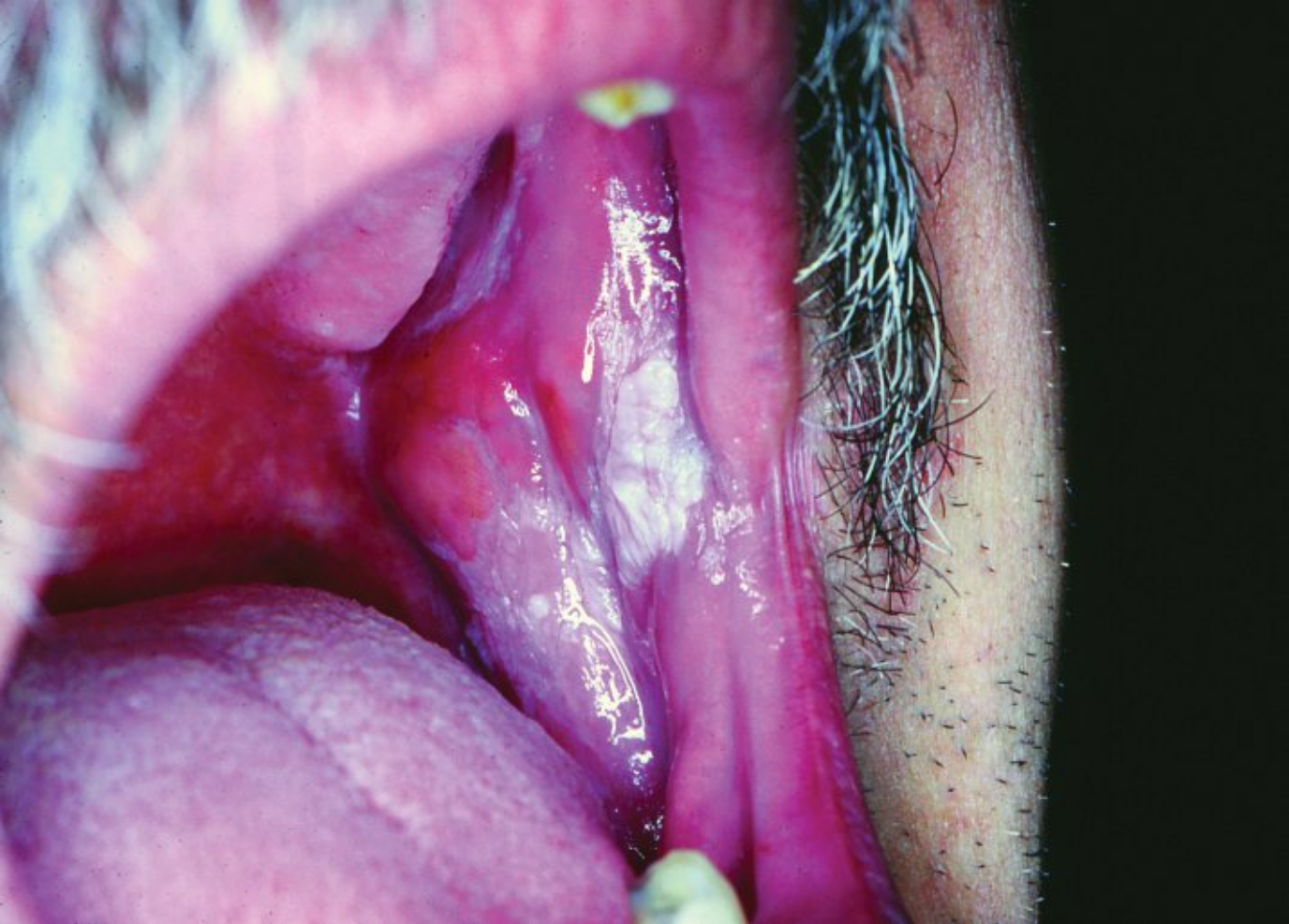
Leukoplakia and Erythroplakia
Leukoplakia is a premalignant lesion that presents as white patches on the oral mucosa (Figure 150). It should be differentiated from oral candidiasis and oral hairy leukoplakia. Oral hairy leukoplakia (Epstein-Barr virus induced) occurs in patients with HIV infection and presents as adherent linear white plaques on the lateral surface of the tongue. Erythroplakia, appearing as red mucosal patches, has a high risk of malignant transformation. Both leukoplakia and erythroplakia require biopsy to rule out dysplasia. These conditions are seen most frequently in tobacco users.
Oral Candidiasis
Oropharyngeal candidiasis presents as white-to-red plaques that are painful (Figure 151). The white surface of the plaques can be scraped off. Candida is also responsible for angular cheilitis (perleche) presenting as fissuring and maceration of the angle of the mouth due to drooling and poorly fitting dentures. Risk factors for oral candidiasis include immunosuppression, diabetes mellitus, use of antibiotics, oral and inhaled glucocorticoids, and smoking. First-line treatment includes azole troches or nystatin swish and swallow mouthwash. Oral fluconazole may be required for severe, recalcitrant, or recurrent cases.
 Oral candidiasis most commonly presents as painful white, fluffy, nonadherent film on the tongue, buccal mucosa, or palate.
Aphthous Ulcers
Aphthous ulcers (“canker sores”) are recurrent ulcers with a gray pseudomembrane base and an erythematous halo that affect lips, buccal mucosa, and tongue (Figure 152). Diagnosis is by clinical findings and history of self-healing. Severe, recurrent oral ulcers suggest the possibility of infection or a systemic disease such as Behçet syndrome, Crohn disease, HIV infection, or erythema multiforme.
Diagnosis of RAS is clinical, and further workup (if any) is aimed at identifying underlying conditions as suggested by the history and examination. On occasion, celiac disease may first present as aphthous ulcers, and so assay for anti-tissue transglutaminase antibodies may be considered. Oral ulcers may also be associated with conditions such as HIV, Crohn disease, and Behçet's syndrome. Topical glucocorticoids are the first-line treatment, but severe RAS that does not respond to steroids can be treated with systemic agents such as colchicine or thalidomide.
Lichen Planus
Oral lichen planus can be isolated or associated with skin lesions or be part of a vulvovaginal syndrome. Oral lesions appear as erythematous patches with an overlying white lacelike pattern (Wickham striae), typically on the buccal mucosa (Figure 153). Treatment often requires systemic steroid-sparing agents in addition to medium- to high-potency topical glucocorticoids.
Actinic Cheilitis and Squamous Cell Carcinoma
Actinic cheilitis is a premalignant condition that presents as scaly patches with erosions on the lower lip and is the result of chronic sun damage. Management is with topical chemotherapy agents (5-fluorouracil), imiquimod, laser therapy, photodynamic therapy, or cryotherapy.
The development of a nodule or ulcer suggests squamous cell carcinoma, most often found on the lower lip of older men (see Common Neoplasms). Risk factors include tobacco, alcohol use, and ultraviolet radiation. Other predisposing conditions include lichen planus and lichen sclerosus. In all cases of suspected squamous cell carcinoma, biopsy is required to establish the diagnosis.
Lichen Sclerosus
Lichen sclerosus is a chronic inflammatory disease that most often affects women in the fifth or sixth decade of life. It presents as a white, atrophic patch that circumferentially involves the vaginal introitus and perianal area with a “figure 8” morphology (Figure 154). Extragenital lesions can occur. Biopsy is required for the diagnosis. Treatment is with potent topical glucocorticoids. If left untreated, permanent scarring can occur, and chronic lesions are at risk for squamous cell carcinoma.
Black Hairy Tongue
Black hairy tongue is characterized by elongation and defective desquamation of filiform papillae (Figure 155). It is more common in men and with advancing age. Management includes good oral hygiene and tongue brushing.
Geographic Tongue
Geographic tongue is fairly common and may be sporadic, familial, or associated with psoriasis. These migratory patterned patches of smooth red surfaces and white patches on the dorsal tongue (Figure 156) are usually asymptomatic, but discomfort and burning may be reported, which are exacerbated by hot or spicy foods. About 40% of patients will develop tongue fissures.