04 GI Tract
typora-copy-images-to: ./images
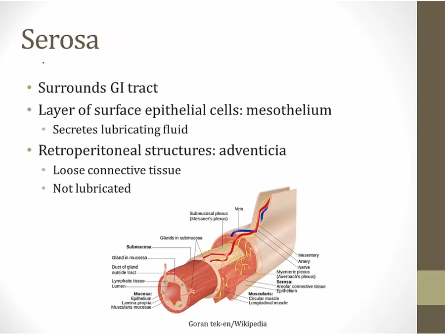
Layers
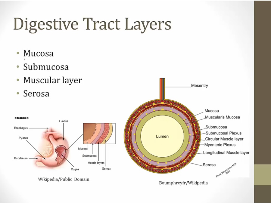
- in all GI tract
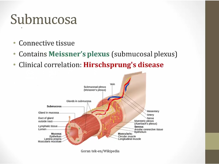
- submucosa nerve plexus
- muscularis nerve plexus
Mucosa
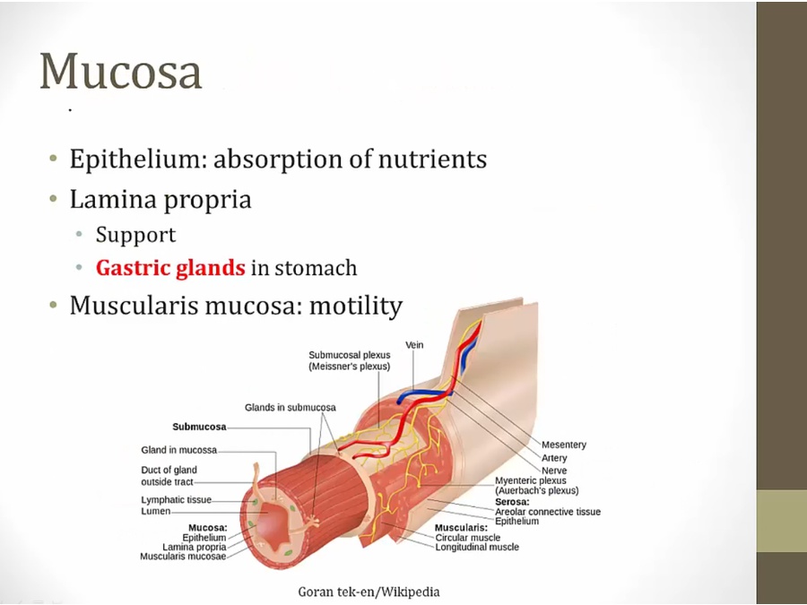
- muscularis within epithelium
Submucosa
Muscular
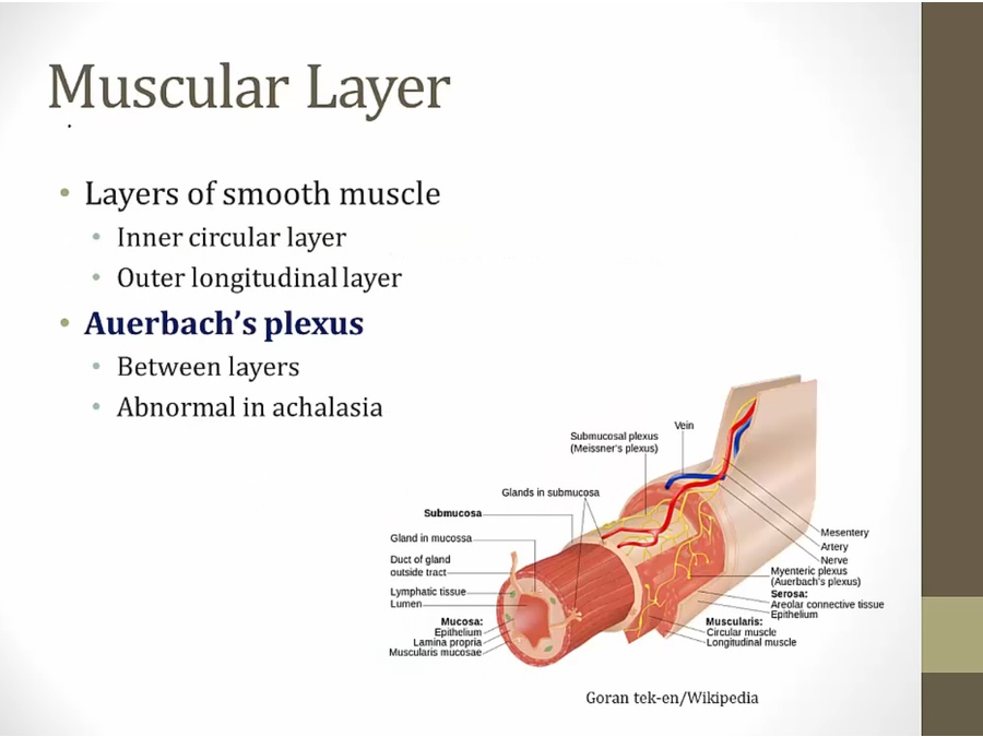
- GI peristalsis
- achalasia: swallowing food problem
Serosa
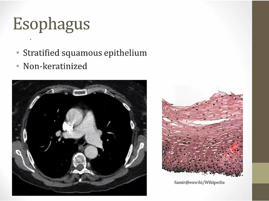
Esophagus
Stomach
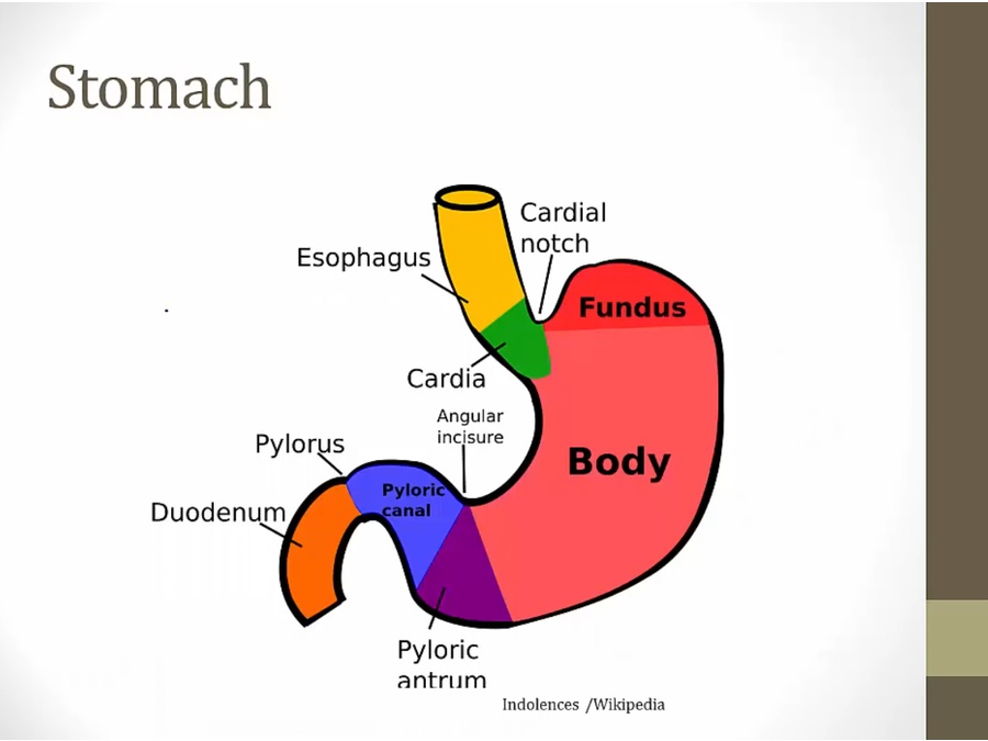
- cardia: connection between esophagus/stomach. Epithelium changes from squamous to columnar
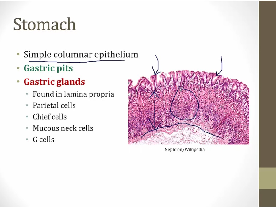
- lamina propria (arrow) between muscular layer (bottom) and epithelium (top)
- gastric glands: within lamina (circle)
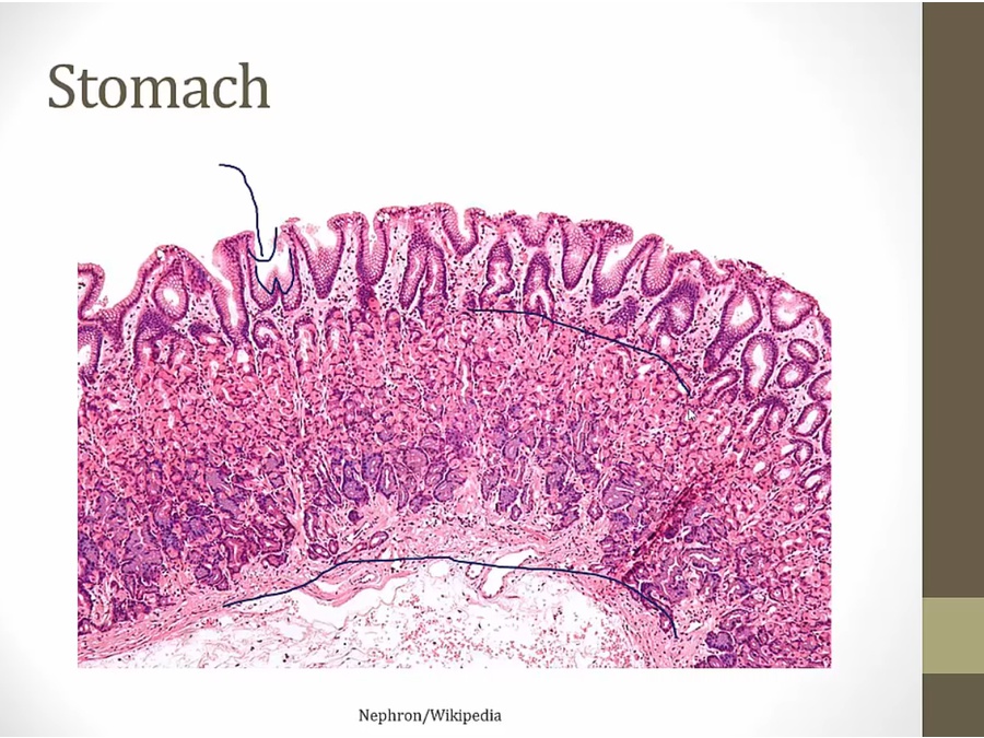
- pink parietal cells near epithelium
- dark blue/purple G cell near bottom
Small intestine
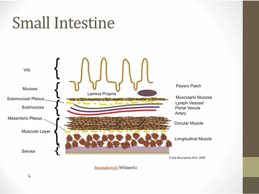
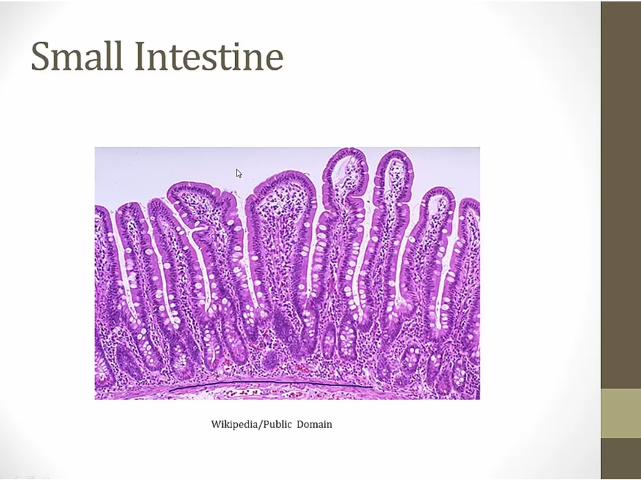
- muscular layer
- columnar epithelium
- only mucosa in villi
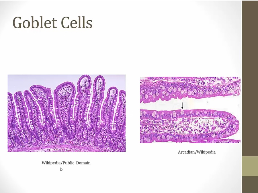
- goblet cell secretes mucous
- microvilli: can't see on light microscope
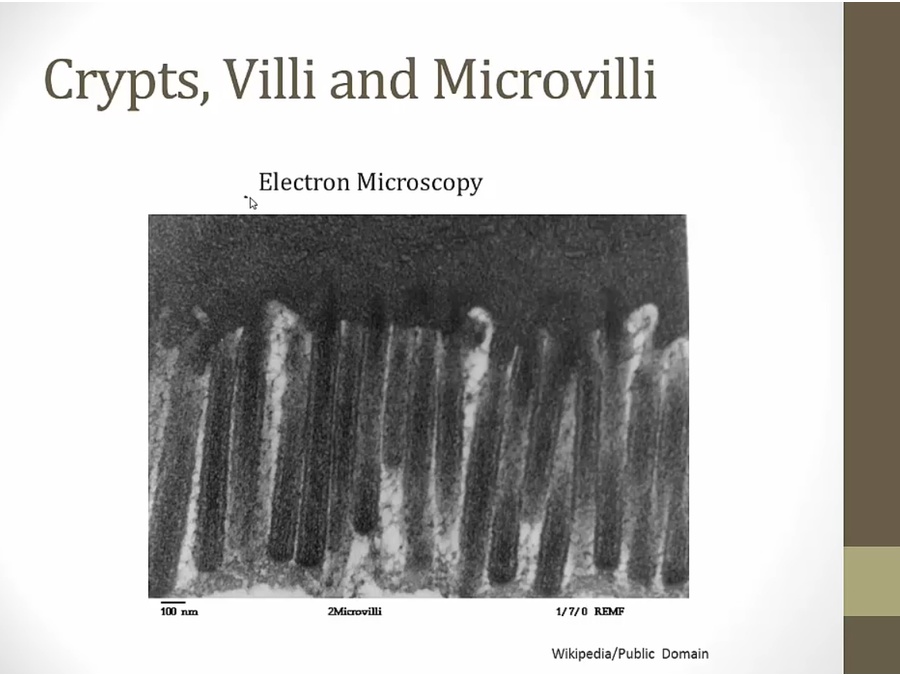
- increase surface areas
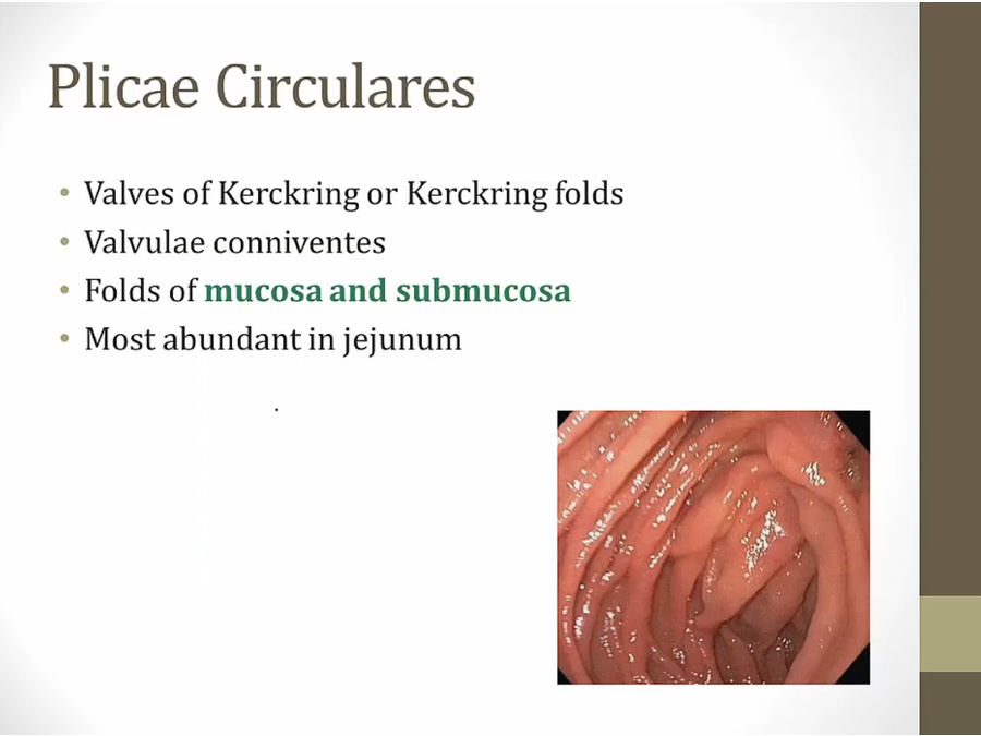
- both mucosa and submucosa
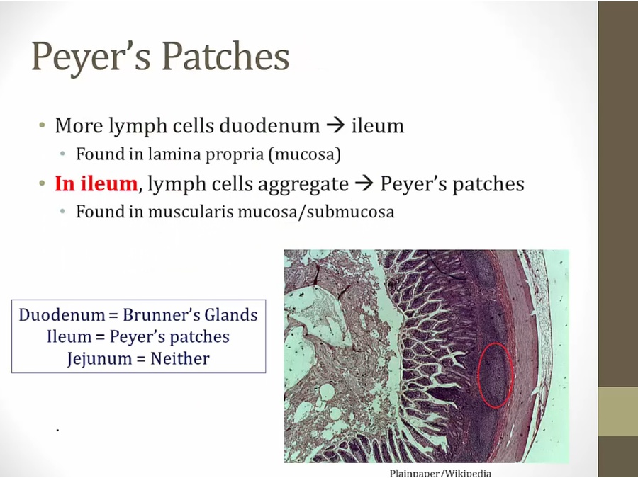
- M cell collect antigens and transmit to Peyer's patch (local lymph)
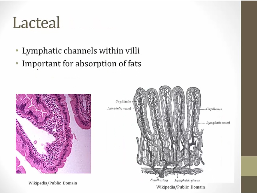
- white space left: lacteal
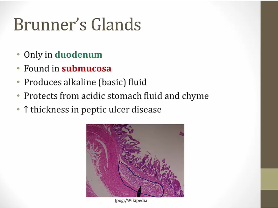
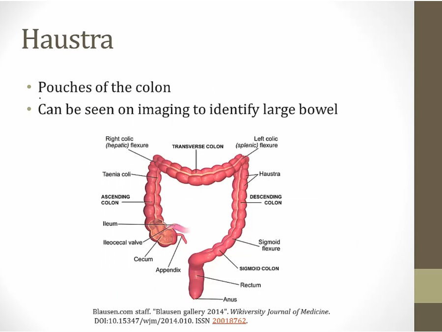
Colon
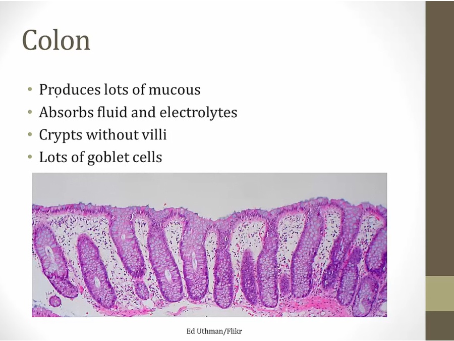
- goblet to produce lots of mucous
Enteric Nervous
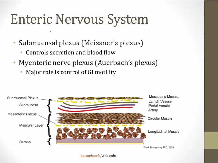
- auerbach between circular and longitudinal muscles
- muscle cells and not neurons
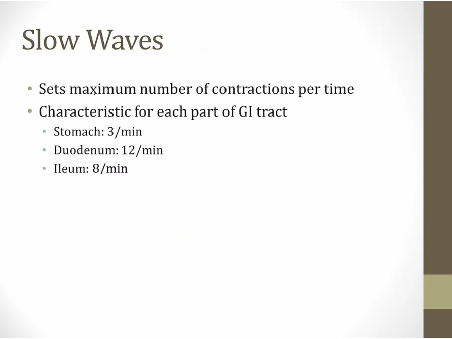
- regulate contractions
- neurons
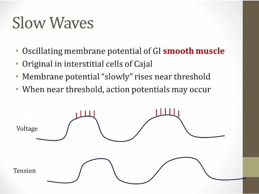
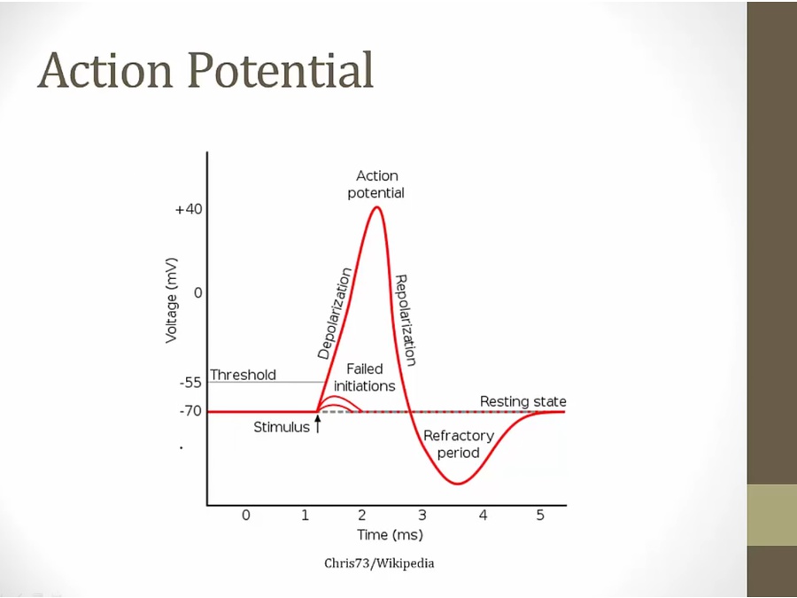
- smooth muscle: resting state fluctuates
Backlinks