01 Cardiac Embryology
Heart contraction
Beginning as early as four weeks gestation.
Primitive heart tube
Formed from Lateral folding directing the endocardial heart tubes to fuse.
The endocardium, myocardium, and epicardium of the heart are derived from mesoderm..
EDC and aorticopulmonary septum derived from ectoderm, neural crest..
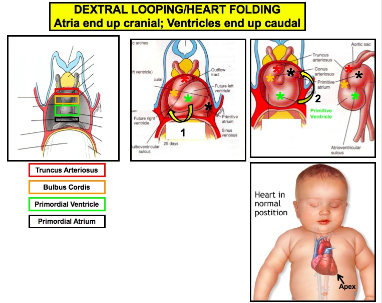
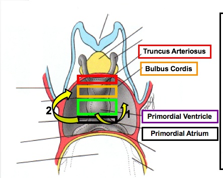
Cardiac looping
begins during the 4th week of gestation, and establishes a left-right polarity to the primitive heart tube. This process requires dynein.
Heart beat
During gestational week 6, transvaginal ultrasound may detect.
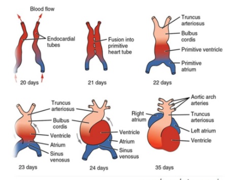
Primitive heart tube
Forms five dilations:
- Truncus arteriosus
- Bulbus cordis
- Primitive ventricle
- Primitive atrium
- **Sinus venosus **(right and left horns).
truncal-bulbar ridge top
left horn/left side: coronary sinus
right horn: RA
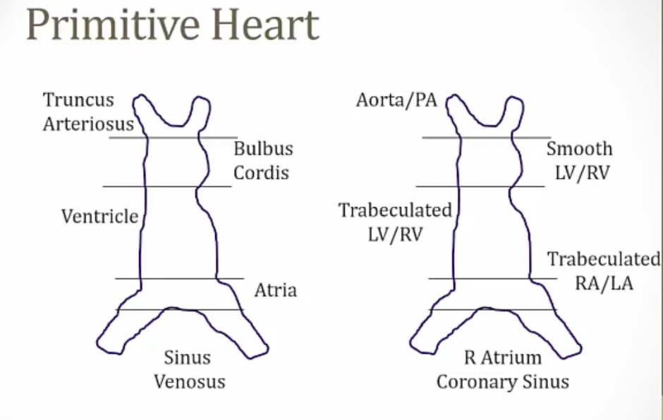
Truncus arteriosus
Develops into the ascending aorta and pulmonary trunk.
Bulbus cordis
Develops into the conus arteriosus (smooth part of the right ventricle) and the aortic vestibule (smooth part of left ventricle).
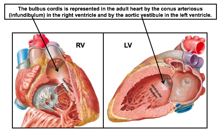
smooth part of right ventricle.
smooth part of left ventricle.
aka conus arteriosus.
Primitive ventricle
Develops into trabeculated part of right and left ventricles.
Primitve atrium
Develops into the muscular (trabeculated) part of right and left atrium and the septum primum.
Left horn of sinus venous
Develops into the coronary sinus. (left side)
Right horn of sinus venous
Develops into the smooth part of the right atrium. (right side)
Crista Terminalis
The junction of the trabeculated and smooth parts of the right atrium.
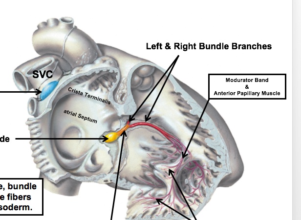
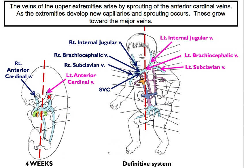
Superior Vena Cava
Structures from outside the heart tube help form the adult heart structure, such as the right common cardinal vein and right anterior cardinal vein which develop into the superior vena cava.
Primitive pulmonary vein
Derives the smooth part of the left atrium.
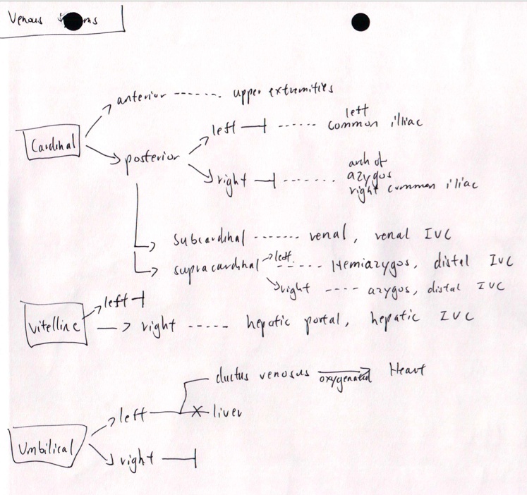
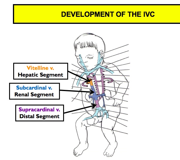
Inferior Vena Cava
Derived from:
- Vitelline veins
- Posterior cardinal veins
- Subcardinal veins
- Supracardinal veins.
Aorticopulmonary septum
Neural crest cells migrate from the hindbrain to the primitive heart tube (Truncal bulbar ridge), which ultimately forms this.
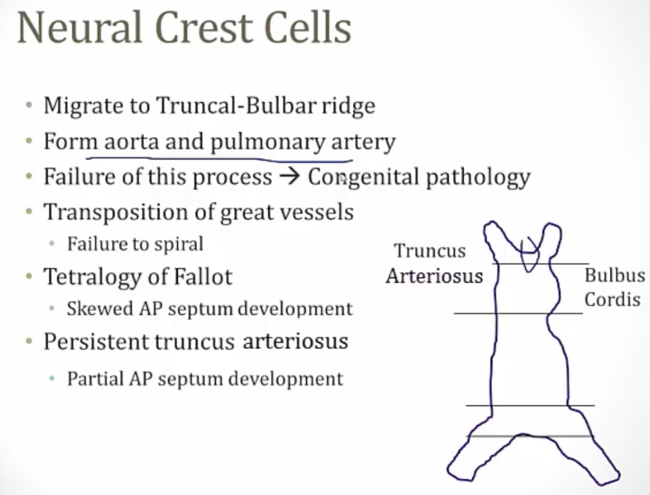
Develops spirally, creating the anterior-posterior relationship between the ascending aorta and pulmonary trunk.
EDC of outflow tract
Derives the aortic and pulmonary valves.
EDC of atrioventricular canal
Derives the mitral and tricuspid valves. Endocardial cushion seprates atria from ventricle.
Heart Septum Formation
Aka ostium primum.
Aka ostium secundum.
Septum Primum
Foramen primum is the first foramen. During the 4th gestational week, a septum separating the primitive atrium begins expanding from the superior interatrial space towards the endocardial cushions, forming septum primum..
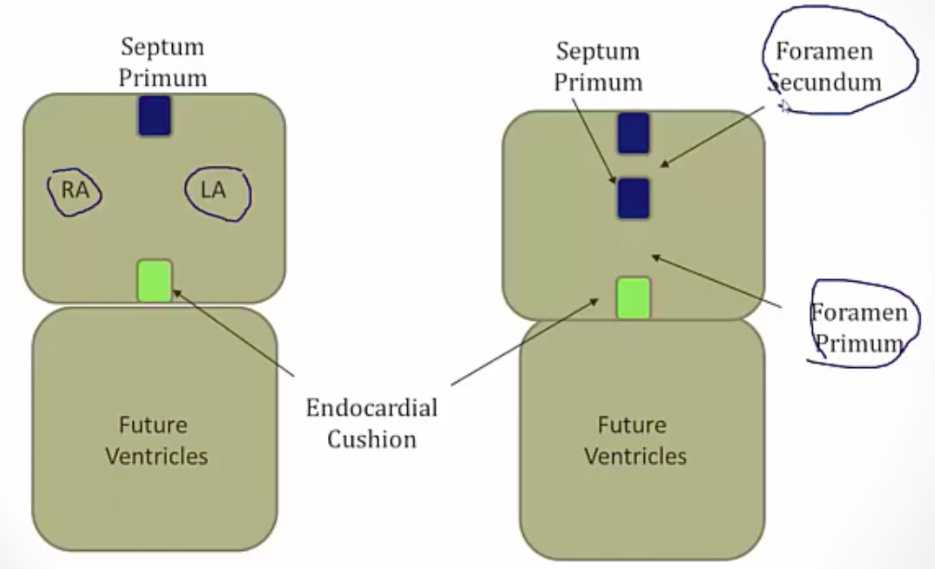
The septum primum does not completely close against the endocardial cushions, leaving the initial opening, foramen primum. The septum primum then develops perforations that join to form foramen secundum. Septum foramen now has two holes in it..
Septum secundum and foramen ovale
The septum secundum grows to the right of the septum primum from top and bottom, forming a partial barrier between the right and left atria. The opening in that barrier is the foramen ovale..
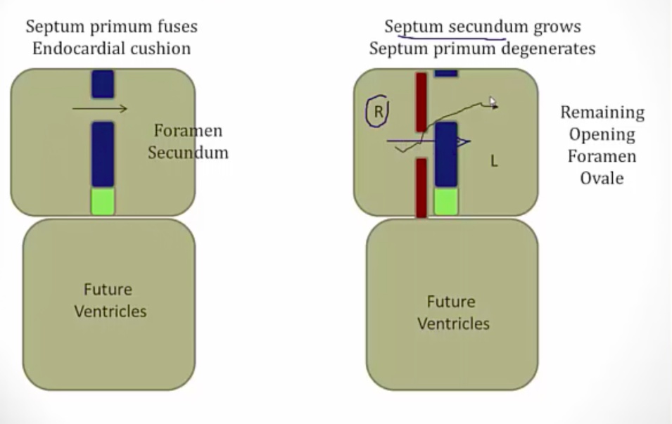
Foramen ovale
The opening in the septum secundum.
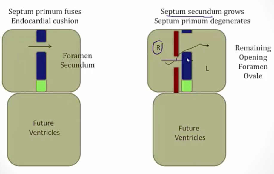
Maintains blood flow from the right atrium to the left atrium.
Fetal Circulation vs Changes in circulation at birth
During fetal circulation, the lungs are filled with fluid (high resistance). Oxygenated blood are carried through umbilical vein and travel straight to RA, bypassing liver via ductus venosus. Blood in RA bypasses lung via foramen ovale. Some blood that goes to RV bypasses lung via ductus arteriosus from PA to aorta..
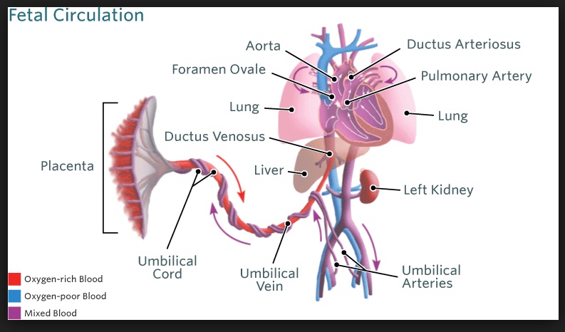
At birth, fluid is pushed out of lung (pulmonary resistance drops). More blood goes to the lungs and thus the left atrium. LA pressure becomes higher than RA. At the same time, closure of umbilical vein decrases RA pressure. This closes the foramen ovale, forming fossa ovalis..
Ductus Arteriosus
Maintained by low O2, high PGE. Low PGE closes ductus..
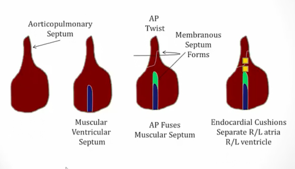
Ventricular septum development
The muscular interventricular septum develops first, originating from the floor of the primitive ventricle and extending towards the endocardial cushions but leaves a space forming the interventricular foramen.
The membranous interventricular septum forms when the **aorticopulmonary septum rotates and fuses with the muscular interventricular septum. **
The membranous IV septum is formed by the fusion of bulbar ridge (part of aorticopulmonary septum) and endocardial cushions
Typically, the rotation of the aorticopulmonary septum and formation of the membranous interventricular septum closes the interventricular foramen..